Glaucoma & Optic Nerve Care

Protect the vision glaucoma can take without warning.

Comprehensive assessment combines eye pressure, drainage-angle examination, optic-nerve imaging and visual-field testing—because no single number can diagnose or exclude glaucoma.

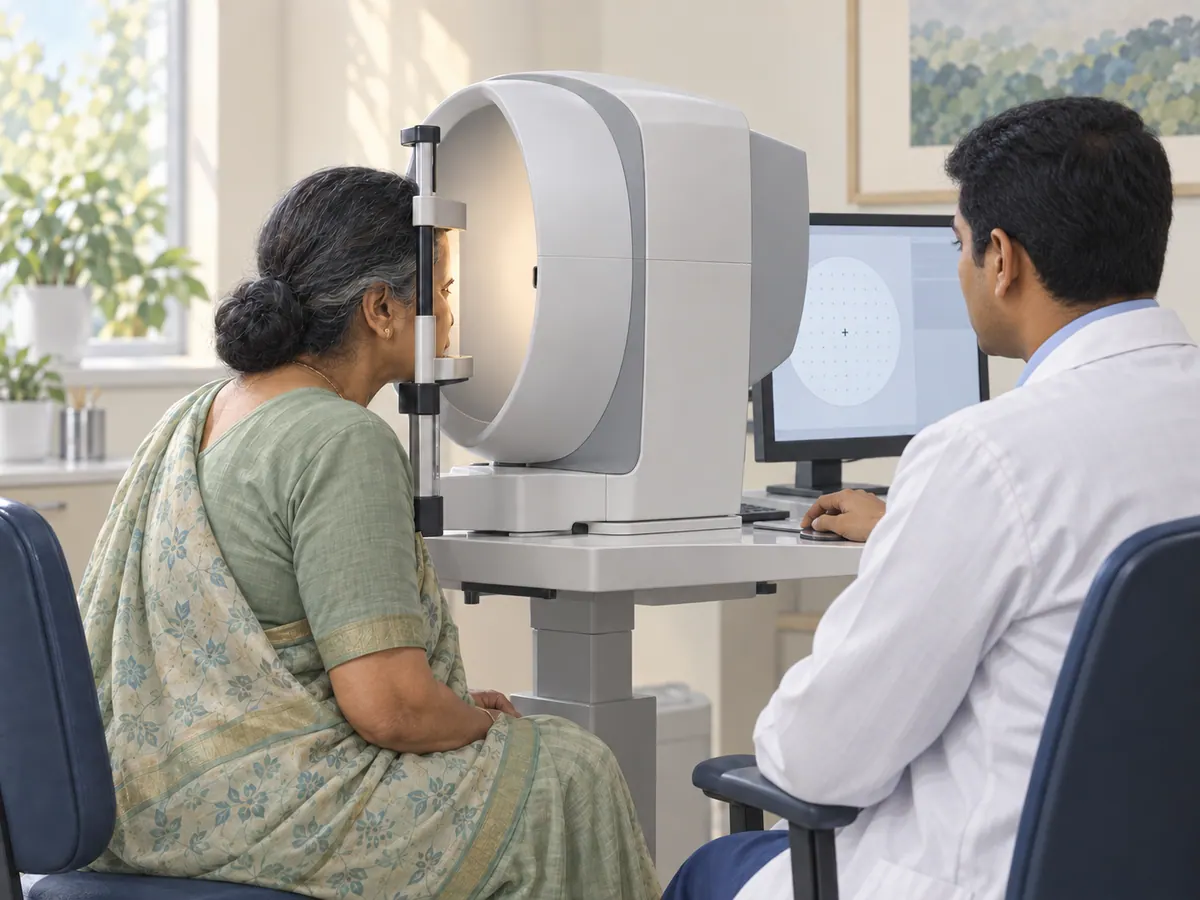
Illustration of a patient undergoing visual field testing for glaucoma
Early detection Protects remaining vision
Understanding glaucoma

Glaucoma is optic-nerve damage—not simply high eye pressure.

The optic nerve carries visual information from the retina to the brain. Glaucoma is a group of diseases in which this nerve becomes progressively damaged, creating permanent blind spots that often begin in side vision.

Eye pressure is the most important modifiable risk factor, but it is only one part of the diagnosis. Some eyes tolerate higher pressure without damage, while others develop glaucoma at pressures within the statistically normal range. The clinician therefore looks for the pattern and progression of nerve injury rather than relying on one pressure cut-off.

Possible acute angle closure

Sudden pain, redness and blur can be an eye emergency.

Most open-angle glaucoma is painless and slow. Acute angle closure is different: pressure can rise rapidly when the drainage angle blocks. Do not wait for a routine clinic appointment if these symptoms appear suddenly.

  • Severe eye pain or a rapidly worsening headache around one eye
  • A red eye with sudden blurred or reduced vision
  • Rainbow-coloured haloes around lights with pain or blur
  • Nausea or vomiting together with eye pain or visual change
  • Sudden marked vision loss after an eye procedure or injury
Types and related conditions

The same pressure reading can mean different things in different eyes.

Classifying the drainage angle and identifying secondary causes affects treatment. “Glaucoma suspect” and ocular hypertension also require thoughtful monitoring, but they do not automatically mean permanent damage is present.

01Common and usually silent

Primary open-angle glaucoma

The drainage angle is physically open, but fluid leaves the eye less efficiently. Optic-nerve damage generally develops slowly, so useful side vision can be lost before a person notices a problem.

How care may be planned

Diagnosis and follow-up combine optic-nerve examination, pressure measurements, OCT and visual-field testing. Treatment is adjusted to an individual target pressure and evidence of progression.

02Narrow or blocked drainage angle

Angle-closure glaucoma

The iris obstructs the eye’s drainage angle. Closure may develop gradually without obvious symptoms, or happen suddenly with severe pain, redness, blurred vision, coloured haloes, headache, nausea or vomiting.

How care may be planned

An acute attack is an emergency. Medicines and laser peripheral iridotomy are commonly used to open an alternative fluid pathway; the other eye may also require preventive assessment or treatment.

03Damage at statistically normal pressure

Normal-tension glaucoma

Characteristic optic-nerve and visual-field damage can occur even when measured eye pressure falls within the usual population range. A single “normal” pressure reading therefore cannot rule out glaucoma.

How care may be planned

The diagnosis requires careful structural and functional assessment and exclusion of other causes of optic-nerve damage. Lowering pressure can still slow progression.

04Raised pressure without proven damage

Ocular hypertension

Some people have higher-than-average eye pressure but no detectable optic-nerve injury or visual-field loss. This is not the same as glaucoma, although it can increase future risk.

How care may be planned

Corneal thickness, pressure level, age, optic-nerve appearance, family history and other risks help decide whether observation or pressure-lowering treatment is appropriate.

05Another condition contributes

Secondary glaucoma

Pseudoexfoliation material, pigment dispersion, inflammation, eye injury, abnormal new vessels, certain medicines or previous eye surgery can interfere with drainage and damage the optic nerve.

How care may be planned

Management addresses the cause where possible while lowering eye pressure. The treatment plan can differ substantially from routine primary open-angle glaucoma care.

06Congenital and paediatric disease

Childhood glaucoma

Rare drainage-development problems can affect babies and children. Warning signs may include excessive tearing, light sensitivity, cloudy corneas or an eye that appears enlarged.

How care may be planned

Prompt specialist evaluation is important because early pressure control supports visual development. Surgery is often central to congenital glaucoma treatment.

Who should be assessed?

Risk rises quietly—family history and a comprehensive exam matter.

Anyone can develop glaucoma. A person with a close family history, suspicious optic nerves, raised pressure, a narrow angle or previous eye injury may need earlier or more frequent examinations. Steroid medicines should never be stopped without the prescribing clinician, but their use should be disclosed during the eye assessment.

Arrange a glaucoma check
  • Increasing age
  • A parent, sibling or child with glaucoma
  • Raised eye pressure or suspicious optic nerves
  • Diabetes, high blood pressure or vascular disease
  • Long-term steroid exposure, including eye drops when medically prescribed
  • Previous eye injury, inflammation or surgery
  • High myopia or other eye-specific anatomical risk
  • A narrow drainage angle identified on examination
Glaucoma diagnostics

Pressure, structure and function must be interpreted together.

Not every test is required at every visit. Good glaucoma care establishes a reliable baseline, then repeats the measurements most useful for deciding whether the condition is stable or progressing.

IOP

Tonometry

Measures pressure inside the eye. Pressure changes through the day and overlaps between healthy and glaucomatous eyes, so a reading above or below 21 mmHg does not diagnose or exclude glaucoma on its own.

ANGLE

Gonioscopy

A mirrored examination lens allows the clinician to inspect the drainage angle and distinguish open-angle, narrow-angle and several secondary forms of glaucoma.

DISC

Dilated optic-nerve examination

The optic-nerve head is assessed for rim thinning, asymmetry, haemorrhage and other features. Photographs can provide a useful baseline for comparison over time.

OCT

Optic-nerve and RNFL OCT

OCT measures the retinal nerve-fibre layer and related structures. Repeat scans help identify structural change, but image quality and the full clinical examination must be considered.

FIELD

Automated perimetry

Visual-field testing maps functional sensitivity and looks for characteristic blind spots. Because attention and technique affect results, reliable baseline and repeat tests are often required.

CCT

Corneal thickness and supporting tests

Pachymetry measures central corneal thickness, which influences interpretation of pressure and risk. Additional tests are selected when the diagnosis or progression remains uncertain.

Monitoring over time

A stable pressure is useful—but stable vision and a stable nerve are the real goal.

Glaucoma decisions are based on trends. The clinician compares pressures, disc appearance, OCT measurements and visual fields with reliable earlier results. Apparent change may need confirmation because scan alignment, cataract, dry eye, concentration and test learning can affect measurements.

A target pressure is an estimate of the range likely to reduce further damage. It can be revised if the nerve or field worsens, if treatment causes problems, or if the overall risk changes.

  1. 01Establish a baseline

    Document the drainage angle, optic nerve, pressure range, corneal thickness, OCT and visual field as indicated.

  2. 02Compare like with like

    Repeat reliable tests at intervals matched to severity and risk.

  3. 03Escalate when needed

    Adjust drops, consider laser or discuss surgery when control or progression requires it.

Treatment choices

Lowering eye pressure is the proven way to slow glaucoma damage.

Choice of treatment depends on glaucoma type, severity, target pressure, previous response, other medical conditions and patient preference. A procedure is not automatically “better” than drops; the aim is reliable long-term control.

01

Pressure-lowering eye drops

Different medicine classes reduce fluid production or improve drainage. The best regimen balances target pressure, other medical conditions, side effects, cost and the ability to use drops consistently.

02

Laser treatment

Selective laser trabeculoplasty may lower pressure in suitable open-angle disease. Laser peripheral iridotomy creates a small opening in the iris for angle-closure risk. These procedures have different purposes and are not interchangeable.

03

Trabeculectomy and drainage surgery

When medicines or laser do not provide adequate control, surgery can create or support a new drainage route. Trabeculectomy and tube-shunt procedures require structured postoperative monitoring.

04

MIGS and combined procedures

Minimally invasive glaucoma procedures may suit selected patients, often with mild-to-moderate open-angle disease or alongside cataract surgery. They are not automatically appropriate for advanced or every type of glaucoma.

The page explains standard treatment categories, not a promise that every procedure is suitable or performed at every JEWCC centre. After examination, the team can recommend medical care, available laser or surgery, or an appropriate referral.

Using glaucoma drops well

The medicine only works when it reaches the eye consistently.

Use the prescribed drop, eye and timing exactly as written. If more than one eye drop is due, leave several minutes between them so the second does not wash out the first. Close the eye gently after instillation; pressing the inner corner beside the nose may reduce medicine drainage into the throat.

Bring every bottle to follow-up. Report redness, wheezing, faintness, unusual fatigue or other concerns rather than stopping treatment independently. Tell the clinician about heart, lung and other medicines because some glaucoma drops have body-wide effects.

At every follow-up
  • Bring current bottles and a medicine list
  • Mention missed doses and practical difficulties honestly
  • Demonstrate technique if you are unsure
  • Ask what to do if a dose is forgotten
  • Keep the next pressure, OCT or visual-field appointment
Insurance and cashless guidance

Testing and procedure benefits may be handled differently.

Many policies distinguish outpatient consultations, OCT and visual-field tests from laser or surgery. Pre-authorisation may require examination findings, test reports, diagnosis, proposed procedure and policy-specific documentation.

JEWCC can assist with available clinical records, but eligibility, consumable limits, co-payments and approval remain the insurer or TPA’s decision.

JEWCC eye centres

Begin with a comprehensive glaucoma assessment near you.

Call the chosen centre before travelling to confirm appointment timing and the availability of specific imaging or visual-field testing. Bring previous prescriptions, pressure records, scans and field reports whenever possible.

Glaucoma FAQs

Clear answers for a long-term condition.

01Can glaucoma be present when eye pressure is normal?

Yes. Normal-tension glaucoma causes characteristic optic-nerve damage despite pressure readings within the usual population range. Conversely, some people have raised pressure without glaucoma. Pressure must be interpreted with the optic nerve, drainage angle, OCT and visual field.

02Is an eye-pressure test enough to detect glaucoma?

No. Tonometry is important, but a single pressure reading can miss glaucoma or incorrectly label a healthy eye. A comprehensive assessment may include gonioscopy, dilated optic-nerve examination, OCT, visual-field testing and corneal-thickness measurement.

03Can vision already lost from glaucoma be restored?

Established glaucomatous optic-nerve damage is generally irreversible. Treatment lowers eye pressure to reduce the risk or rate of further loss. This is why early detection and reliable follow-up matter even when vision feels normal.

04Will glaucoma eye drops be needed for life?

Glaucoma is usually a long-term condition. Drops often continue unless the clinician changes the regimen or laser or surgery provides sufficient control. Never stop a prescribed drop because the eye feels comfortable; discuss side effects or cost so an alternative can be considered.

05How often are OCT and visual-field tests repeated?

The interval depends on risk, disease severity, reliability of earlier tests, recent treatment and suspected progression. New or unstable glaucoma may need closer testing, while stable disease may be reviewed less frequently. The clinician sets an individual schedule.

06Does a family history mean I will develop glaucoma?

No, but a close relative with glaucoma increases risk. Tell the clinician which relative was affected and, if known, the type and age at diagnosis. Regular comprehensive eye examinations can detect pressure or optic-nerve changes before symptoms develop.

07Are glaucoma tests, laser or surgery covered by insurance?

Coverage varies by policy. Outpatient consultations, visual fields and OCT may have different rules from laser or surgery. Procedures may need pre-authorisation, investigation reports and a confirmed diagnosis. JEWCC can help prepare available documents, while the insurer or TPA makes the final decision.